Dermatochalasis, a term that may sound complex, refers to the excess skin that can appear on our upper and/or lower eyelids. As we age, our skin, especially the delicate skin around our eyes, undergoes various changes. This article will get you into the world of dermatochalasis, breaking down its symptoms, causes, and the best ways to manage it.
Whether you’re a concerned individual or just curious, this guide is designed to provide you with comprehensive insights in an easy-to-understand format.
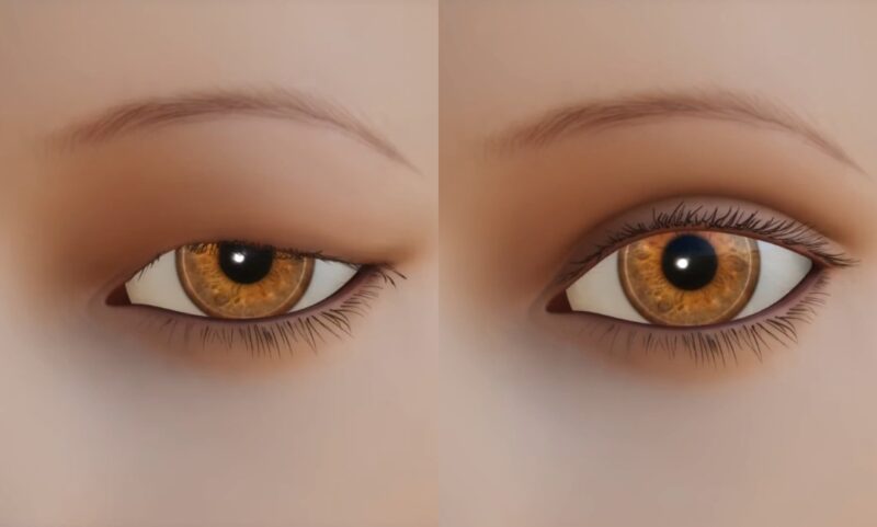
Dermatochalasis is characterized by excess skin on the upper and/or lower eyelids. This condition is commonly associated with aging, but it can also affect younger adults.
Characteristics of Dermatochalasis
The eyelid skin is notably thin, making it susceptible to fine wrinkling. As time progresses, various factors contribute to the development of redundant skin. This can be accompanied by:
- Fat herniation, especially in the superomedial region and in the inferomedial and central part.
- Descent of eyebrows and mid-face.
- Puffiness due to eyelid edema.
- Discoloration from venous leakage, leading to dark circles under the eyes.
Common Contributing Factors
Several factors play a role in the development of this lax and redundant eyelid tissue:
- Aging: As we grow older, our skin naturally loses its elasticity.
- Loss of Elastic Tissue: The skin’s elastic tissue diminishes over time.
- Weakening of Connective Tissue: The eyelid’s connective tissue becomes weaker.
- Gravity’s Pull: The constant downward force of gravity affects our skin.
- Solar and Age-Related Degeneration: Exposure to the sun and natural aging can degrade collagen.
- Descent of Facial Complexes: This includes the descent of the brow complex (ROOF) and malar complex (SOOF).
- Other Factors: Actinic exposure and smoking can accelerate the development of these changes.
Symptoms of Dermatochalasis
While dermatochalasis can sometimes be asymptomatic, it usually presents bilaterally, affecting both eyes.
Common Symptoms
Individuals with dermatochalasis might experience:
- Redundant upper eyelid skin, possibly with herniation of orbital fat.
- Obstruction of superior visual fields due to excess skin.
- Eye irritation and blepharitis.
- Ectropion of the lower eyelid or entropion of the upper eyelid.
- Dermatitis and a feeling of fullness or heaviness in the upper eyelids.
- “Bags” appearing in the lower eyelids.
- Wrinkles in the lower eyelid and at the lateral canthus.
Causes Behind the Symptoms
The primary cause of dermatochalasis is age-related loss of skin elasticity and weakening of the eyelid’s connective tissue. This condition is predominantly seen in the elderly.
Diagnosis

Understanding dermatochalasis requires a comprehensive diagnosis based on clinical features.
Pathophysiology and Histology
The pathophysiology of dermatochalasis aligns with the normal aging changes of the eyelid skin. This encompasses:
- Loss of elastic fibers.
- Thinning of the epidermis.
- Redundancy of the skin.
- Presence of chronic infiltrate when associated with dermatitis.
Histologically, the epidermis appears thin and smooth. The dermis may show some loss of elastic and collagen tissue, increased capillary vascularity, and basophilic degeneration of collagen.
Systemic Associations
Certain systemic diseases can predispose individuals to develop dermatochalasis. These include:
- Thyroid eye disease.
- Hereditary angioneurotic edema.
- Trauma and renal disease.
- Conditions like amyloidosis, cutis laxa, and Ehlers-Danlos syndrome.
- Xanthelasma, with genetic factors playing a role in some cases.
Comprehensive Evaluation

Before diving into treatment options, a thorough evaluation is essential to ensure the best possible outcomes.
Key Evaluation Steps
When evaluating dermatochalasis, medical professionals look for:
- Pre-existing conditions such as blepharoptosis, brow ptosis, and lacrimal gland prolapse.
- Lateral bulging of the upper lid, which may indicate lacrimal gland prolapse.
- Coexistent dermatochalasis of the eyelids and browptosis. This can be tested by elevating the eyebrows.
- Measurements of the interpalpebral fissure, levator function, and the marginal reflex distance from both the upper and lower eyelids.
- The eyelid crease should also be measured in millimeters.
Differential Diagnosis
Dermatochalasis can sometimes be confused with other conditions. It’s crucial to differentiate it from:
- Congenital or acquired ptosis.
- Eyebrow ptosis.
- Floppy eyelid syndrome.
- Eyelid oedema or blepharochalasis.
- Prolapse of the lacrimal gland.
Be careful as in cases your lacrimal gland can get inflamed and cause disease called dacryoadenitis, early detection and proper treatment are essential
Management Strategies
Managing dermatochalasis often requires a combination of medical and surgical interventions.
Medical Therapy
While surgery is the most definitive treatment, certain medical therapies can alleviate symptoms:
- Lid Hygiene: Essential for those suffering from blepharitis.
- Topical Antibiotics: Can be prescribed for infections.
- Topical Steroids: Beneficial for those with dermatitis.
- Lubricants: These can provide relief for patients with dry eyes.
Surgical Therapy
For many, surgical intervention offers the best chance at symptom relief:
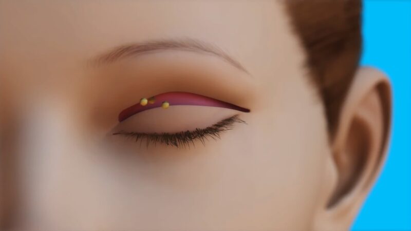
- Upper Eyelid Blepharoplasty: This procedure removes excess eyelid skin and orbital fat, reconstructing the upper eyelid crease.
- Ptosis Surgery: Beneficial for patients with associated ptosis or drooping of the eyelid.
- Other Procedures: These include the placement of temporary collagen punctal plugs, permanent punctal plugs, or punctal cautery for those with dry eyes.
Prognosis and Complications
Understanding the potential outcomes and risks is crucial for anyone considering treatment for dermatochalasis.
Prognosis
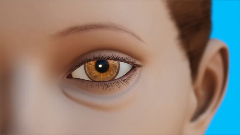
With accurate diagnosis and appropriate surgical intervention, the results are generally positive. Surgical blepharoplasty, in particular, has shown to offer significant improvements for patients.
Potential Complications
As with any medical condition or procedure, there are potential complications to be aware of:
- Lagophthalmos or the inability to close the eyes completely.
- Ptosis or drooping of the upper eyelid.
- Keratitis, an inflammation of the cornea.
- Eyelid retraction.
- Conjunctival chemosis, or swelling of the conjunctiva.
FAQ
Can dermatochalasis affect both eyes simultaneously?
Yes, dermatochalasis typically presents bilaterally, meaning it affects both eyes at the same time. However, the severity can vary between the two eyes.
Is dermatochalasis the same as having “baggy eyes”?
While “baggy eyes” can be a symptom of dermatochalasis due to fat herniation and skin redundancy, not everyone with baggy eyes has dermatochalasis. Other factors like fluid retention, allergies, or lack of sleep can also cause baggy eyes.
Can lifestyle changes prevent or reduce the severity of dermatochalasis?
While aging and genetic factors play a significant role, avoiding smoking, reducing sun exposure, and maintaining good skin care can potentially delay or reduce the severity of dermatochalasis.
Is the surgery for dermatochalasis purely cosmetic?
No, while many people opt for surgery due to cosmetic concerns, the excess skin from dermatochalasis can obstruct vision, making surgery a functional necessity for some.
How long is the recovery period after surgery?
Recovery can vary, but most patients can return to their regular activities within 10-14 days post-surgery. Complete healing and settling of scars might take a few months.
Final Words
Dermatochalasis, though a common condition, often remains misunderstood. Whether you’re exploring treatment options or seeking to understand the changes in your skin, knowledge is empowering. Remember, every individual’s experience with dermatochalasis is unique. Consultation with a medical professional can provide tailored advice and solutions. Stay informed, and take proactive steps towards eye health and overall well-being.